Science outreach
At NCCAT and NYSBC we outreach to the community to provide first-hand exposure to microscopy techniques and disseminate information on how we as structural biologists understand molecular machines (biological complexes that carry out multiple functions within our
What is cryo-electron microscopy?
How does cryoEM help biomedical research?
Public Health Relevance
The structure of a molecule reveals important information about how it functions and can help scientists identify potential new therapeutic targets for vaccines and drugs to combat diseases and conditions. Cryo-electron microscopy (cryo-EM) is a method used to image frozen biological molecules, such as proteins and nucleic acids, without the need for structure-altering dyes or crystallization. Not only can cryo-EM image molecules in their natural shapes, but it can obtain structures of molecules that were impossible using other methods like X-ray crystallography. Recent advances in cryo-EM methods and technology have greatly extended its resolution and the amount of information it can provide about viruses, proteins, and other important biomolecules.
Find out more here: https://commonfund.nih.gov/cryoem/.
Outreach events and materials
SEMC calendar
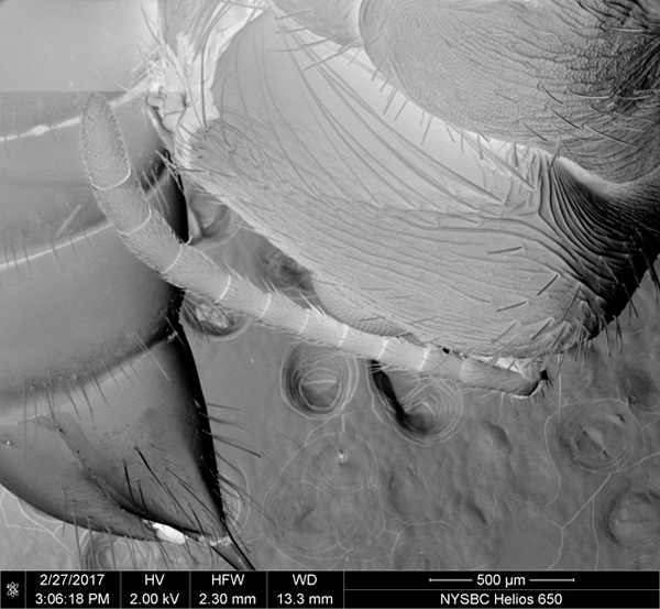
Looking at everyday objects up close using a scanning electron microscope (SEM) (with images by M. Kopylov).
Math for America
Helping teachers learn about the advanced techniques of structural biology at NYSBC including NMR Spectroscopy, X-ray crystallography, cryo-electron microscopy. Educators have the opportunity to see the equipment first-hand.
Guggenheim
The Hugo Boss Prize 2016 winner Anicka Yi’s Life Is Cheap exhibit opening reception was held on Thursday, April 20, 2017. For their invitation, they used a scanning electron microscope (SEM) image of an ant taken at NYSBC.